
Shave biopsy
A thin layer of the spot is removed with a small blade. Often used for raised or surface lesions.
Skin Cancer Center · Biopsy & Pathology
A skin biopsy is how a skin cancer diagnosis is confirmed. Your pathology report names the exact diagnosis and the features that guide treatment — and at Juniper, that report drives a clear, pathology-guided plan explained in plain language.
Medically reviewed by Dr. Karla Snider Pivik, DO, FAAD, FAOCD — board‑certified dermatologist
The short answer
A biopsy removes a small sample of skin so a pathologist can examine it under a microscope. It is the step that confirms whether a spot is a basal cell carcinoma, a squamous cell carcinoma, a melanoma, a precancer, or something benign. Because treatment is different for each, an exact diagnosis comes first — then the plan follows the report.
✓ A skin biopsy removes a small sample of skin for a pathologist to examine
✓ It is the way a skin cancer diagnosis is confirmed
✓ Your pathology report names the diagnosis and features that guide treatment
✓ At Juniper, the report drives a dermatologist-led, pathology-guided plan
Types of biopsy
The technique depends on the spot’s size, location, and what the dermatologist needs to evaluate.
A thin layer of the spot is removed with a small blade. Often used for raised or surface lesions.
A small circular tool removes a deeper core of skin, which can help evaluate deeper features.
All or part of a larger lesion is removed with a scalpel when a deeper or complete sample is needed.
Reading your report
Reports use clinical language. Here is what the common sections describe — and we will always walk through your specific report with you.
The specific finding — for example basal cell carcinoma, squamous cell carcinoma, melanoma, a precancer, or a benign spot.
Describes how the cells look, which can affect how a cancer is expected to behave and how it is treated.
Whether cancer cells reach the edge of the sample. This helps determine whether more tissue needs to be removed.
How deep the abnormal cells extend. For melanoma this is often reported as Breslow thickness and guides next steps.
Findings such as perineural involvement or certain growth patterns can raise risk and change the recommendation.
Pathology-guided care
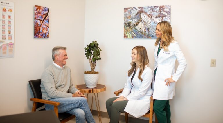
A pathology report can feel intimidating. Our job is to translate it — what the diagnosis means, what the margins and other features tell us, and what happens next. From there we build a plan that fits your diagnosis, its location, your health, and your goals.
Juniper brings skin checks, biopsy, pathology-guided planning, and surgical excision together in Bend and Redmond, with GentleCure® Image-Guided SRT available in Bend for selected nonmelanoma cancers.
Steps to answers
Why choose Juniper
Waiting on biopsy results is stressful. We aim to review results promptly and explain them honestly, so you understand your diagnosis and your options — without a long, anxious wait.
Plain-language explanations. We walk through your report line by line so the diagnosis and next steps are clear.
Pathology-guided planning. Treatment recommendations follow the report — not a one-size-fits-all script.
Local, coordinated care. Skin checks, biopsy, pathology-guided planning, and surgical excision are offered in Bend and Redmond.
Timely follow-up. We aim to review results promptly so you are not left waiting for answers.
Questions & answers
A skin biopsy is a quick, in-office procedure in which a small sample of skin is removed and sent to a pathologist. Examining the tissue under a microscope is how a skin cancer diagnosis is confirmed and how the specific type is identified.
The area is numbed with a local anesthetic first, so most people feel a brief sting from the numbing and little else during the biopsy. Your provider will review aftercare for the small wound.
Turnaround depends on the pathology lab and whether additional testing is needed. Your Juniper care team will tell you how results are communicated and when to follow up if you have not heard from us.
Margins describe whether abnormal cells reach the edge of the removed sample. If the margins are not clear, your dermatologist may recommend removing more tissue to reduce the chance the cancer was left behind. We will explain what your specific margins mean for your plan.
We review your pathology report with you in plain language and outline the appropriate options based on the diagnosis, its location, and your health. From there we build a pathology-guided plan together — which may include monitoring, surgical excision, coordinated specialty care, or GentleCure® for selected nonmelanoma cancers.
Not necessarily. Some biopsies sample only part of a lesion to confirm the diagnosis, so treatment may still be needed even if the biopsy site has healed. Your report and your dermatologist's exam determine whether more treatment is recommended.
Your biopsy is read by a pathologist, and your results and plan are reviewed with a Juniper dermatology provider. Depending on availability and your appointment type, that may be Dr. Karla Pivik or another qualified member of the team.
Independent sources
These independent, authoritative resources can help you understand your biopsy and report.
Bend & Redmond · Central Oregon
Dermatologist-led skin cancer evaluation and pathology-guided planning in Bend and Redmond.